Molybdenum »
PDB 1n62-2c9x »
1vlb »
Molybdenum in PDB 1vlb: Structure Refinement of the Aldehyde Oxidoreductase From Desulfovibrio Gigas at 1.28 A
Enzymatic activity of Structure Refinement of the Aldehyde Oxidoreductase From Desulfovibrio Gigas at 1.28 A
All present enzymatic activity of Structure Refinement of the Aldehyde Oxidoreductase From Desulfovibrio Gigas at 1.28 A:
1.2.3.1;
1.2.3.1;
Protein crystallography data
The structure of Structure Refinement of the Aldehyde Oxidoreductase From Desulfovibrio Gigas at 1.28 A, PDB code: 1vlb
was solved by
J.M.Rebelo,
J.M.Dias,
R.Huber,
J.J.G.Moura,
M.J.Romao,
with X-Ray Crystallography technique. A brief refinement statistics is given in the table below:
| Resolution Low / High (Å) | 24.40 / 1.28 |
| Space group | P 61 2 2 |
| Cell size a, b, c (Å), α, β, γ (°) | 141.780, 141.780, 160.870, 90.00, 90.00, 120.00 |
| R / Rfree (%) | 14.8 / 19.3 |
Other elements in 1vlb:
The structure of Structure Refinement of the Aldehyde Oxidoreductase From Desulfovibrio Gigas at 1.28 A also contains other interesting chemical elements:
| Magnesium | (Mg) | 2 atoms |
| Iron | (Fe) | 4 atoms |
| Chlorine | (Cl) | 3 atoms |
Molybdenum Binding Sites:
The binding sites of Molybdenum atom in the Structure Refinement of the Aldehyde Oxidoreductase From Desulfovibrio Gigas at 1.28 A
(pdb code 1vlb). This binding sites where shown within
5.0 Angstroms radius around Molybdenum atom.
In total only one binding site of Molybdenum was determined in the Structure Refinement of the Aldehyde Oxidoreductase From Desulfovibrio Gigas at 1.28 A, PDB code: 1vlb:
In total only one binding site of Molybdenum was determined in the Structure Refinement of the Aldehyde Oxidoreductase From Desulfovibrio Gigas at 1.28 A, PDB code: 1vlb:
Molybdenum binding site 1 out of 1 in 1vlb
Go back to
Molybdenum binding site 1 out
of 1 in the Structure Refinement of the Aldehyde Oxidoreductase From Desulfovibrio Gigas at 1.28 A
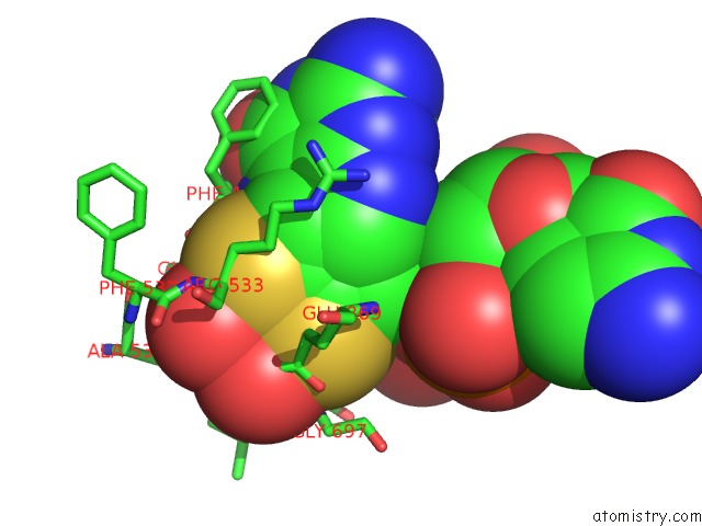
Mono view
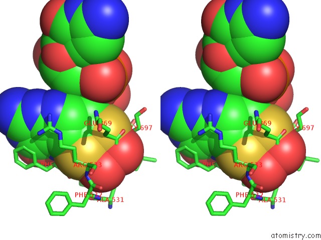
Stereo pair view
Mono view
Stereo pair view
A full contact list of Molybdenum with other atoms in the Mo binding
site number 1 of Structure Refinement of the Aldehyde Oxidoreductase From Desulfovibrio Gigas at 1.28 A within 5.0Å range:
|
Reference:
J.M.Rebelo,
J.M.Dias,
R.Huber,
J.J.G.Moura,
M.J.Romao.
Structure Refinement of the Aldehyde Oxidoreductase From Desulfovibrio Gigas (Mop) at 1.28 A J.Biol.Inorg.Chem. V. 6 791 2001.
ISSN: ISSN 0949-8257
PubMed: 11713686
DOI: 10.1007/S007750100255
Page generated: Sun Aug 17 03:02:53 2025
ISSN: ISSN 0949-8257
PubMed: 11713686
DOI: 10.1007/S007750100255
Last articles
K in 9NESK in 9PHG
K in 9NEI
K in 9NED
K in 9NEC
K in 9NEG
K in 9CWU
K in 9CVB
K in 9CVA
K in 9COM