Molybdenum »
PDB 2ca3-3fah »
2iv2 »
Molybdenum in PDB 2iv2: Reinterpretation of Reduced Form of Formate Dehydrogenase H From E. Coli
Enzymatic activity of Reinterpretation of Reduced Form of Formate Dehydrogenase H From E. Coli
All present enzymatic activity of Reinterpretation of Reduced Form of Formate Dehydrogenase H From E. Coli:
1.17.1.9; 1.17.99.7;
1.17.1.9; 1.17.99.7;
Protein crystallography data
The structure of Reinterpretation of Reduced Form of Formate Dehydrogenase H From E. Coli, PDB code: 2iv2
was solved by
H.C.A.Raaijmakers,
M.J.Romao,
with X-Ray Crystallography technique. A brief refinement statistics is given in the table below:
| Resolution Low / High (Å) | 34.50 / 2.27 |
| Space group | P 41 21 2 |
| Cell size a, b, c (Å), α, β, γ (°) | 146.400, 146.400, 81.270, 90.00, 90.00, 90.00 |
| R / Rfree (%) | 17.5 / 22.6 |
Other elements in 2iv2:
The structure of Reinterpretation of Reduced Form of Formate Dehydrogenase H From E. Coli also contains other interesting chemical elements:
| Iron | (Fe) | 4 atoms |
Molybdenum Binding Sites:
The binding sites of Molybdenum atom in the Reinterpretation of Reduced Form of Formate Dehydrogenase H From E. Coli
(pdb code 2iv2). This binding sites where shown within
5.0 Angstroms radius around Molybdenum atom.
In total only one binding site of Molybdenum was determined in the Reinterpretation of Reduced Form of Formate Dehydrogenase H From E. Coli, PDB code: 2iv2:
In total only one binding site of Molybdenum was determined in the Reinterpretation of Reduced Form of Formate Dehydrogenase H From E. Coli, PDB code: 2iv2:
Molybdenum binding site 1 out of 1 in 2iv2
Go back to
Molybdenum binding site 1 out
of 1 in the Reinterpretation of Reduced Form of Formate Dehydrogenase H From E. Coli
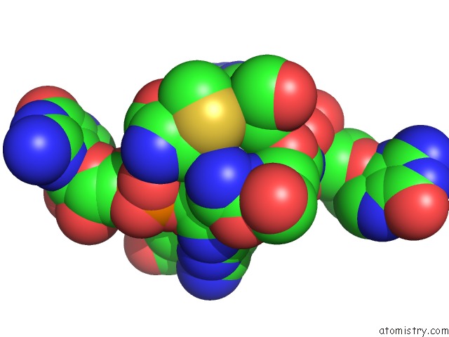
Mono view
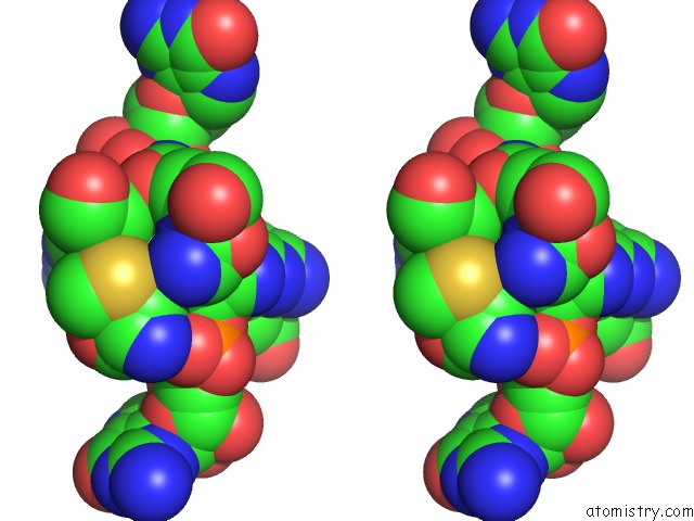
Stereo pair view
Mono view
Stereo pair view
A full contact list of Molybdenum with other atoms in the Mo binding
site number 1 of Reinterpretation of Reduced Form of Formate Dehydrogenase H From E. Coli within 5.0Å range:
|
Reference:
H.C.A.Raaijmakers,
M.J.Romao.
Formate-Reduced E. Coli Formate Dehydrogenase H: the Reinterpretation of the Crystal Structure Suggests A New Reaction Mechanism. J.Biol.Inorg.Chem. V. 11 849 2006.
ISSN: ISSN 0949-8257
PubMed: 16830149
DOI: 10.1007/S00775-006-0129-2
Page generated: Sun Oct 6 15:46:02 2024
ISSN: ISSN 0949-8257
PubMed: 16830149
DOI: 10.1007/S00775-006-0129-2
Last articles
Zn in 9J0NZn in 9J0O
Zn in 9J0P
Zn in 9FJX
Zn in 9EKB
Zn in 9C0F
Zn in 9CAH
Zn in 9CH0
Zn in 9CH3
Zn in 9CH1