Molybdenum »
PDB 4c80-5koj »
4uhx »
Molybdenum in PDB 4uhx: Human Aldehyde Oxidase in Complex with Phthalazine and Thioridazine
Enzymatic activity of Human Aldehyde Oxidase in Complex with Phthalazine and Thioridazine
All present enzymatic activity of Human Aldehyde Oxidase in Complex with Phthalazine and Thioridazine:
1.2.3.1;
1.2.3.1;
Protein crystallography data
The structure of Human Aldehyde Oxidase in Complex with Phthalazine and Thioridazine, PDB code: 4uhx
was solved by
C.Coelho,
M.J.Romao,
T.Santos-Silva,
with X-Ray Crystallography technique. A brief refinement statistics is given in the table below:
| Resolution Low / High (Å) | 105.17 / 2.70 |
| Space group | P 42 21 2 |
| Cell size a, b, c (Å), α, β, γ (°) | 148.732, 148.732, 132.808, 90.00, 90.00, 90.00 |
| R / Rfree (%) | 19.526 / 24.408 |
Other elements in 4uhx:
The structure of Human Aldehyde Oxidase in Complex with Phthalazine and Thioridazine also contains other interesting chemical elements:
| Iron | (Fe) | 4 atoms |
Molybdenum Binding Sites:
The binding sites of Molybdenum atom in the Human Aldehyde Oxidase in Complex with Phthalazine and Thioridazine
(pdb code 4uhx). This binding sites where shown within
5.0 Angstroms radius around Molybdenum atom.
In total only one binding site of Molybdenum was determined in the Human Aldehyde Oxidase in Complex with Phthalazine and Thioridazine, PDB code: 4uhx:
In total only one binding site of Molybdenum was determined in the Human Aldehyde Oxidase in Complex with Phthalazine and Thioridazine, PDB code: 4uhx:
Molybdenum binding site 1 out of 1 in 4uhx
Go back to
Molybdenum binding site 1 out
of 1 in the Human Aldehyde Oxidase in Complex with Phthalazine and Thioridazine
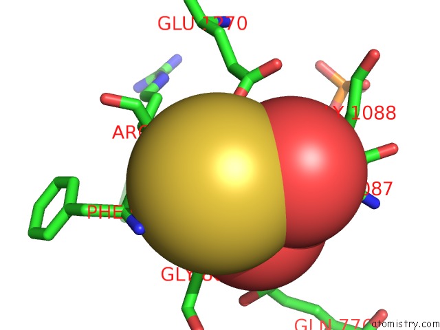
Mono view
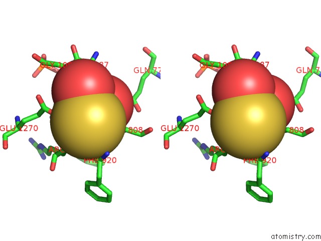
Stereo pair view
Mono view
Stereo pair view
A full contact list of Molybdenum with other atoms in the Mo binding
site number 1 of Human Aldehyde Oxidase in Complex with Phthalazine and Thioridazine within 5.0Å range:
|
Reference:
C.Coelho,
A.Foti,
T.Hartmann,
T.Santos-Silva,
S.Leimkuhler,
M.J.Romao.
Structural Insights Into Xenobiotic and Inhibitor Binding to Human Aldehyde Oxidase Nat.Chem.Biol. V. 11 779 2015.
ISSN: ISSN 1552-4450
PubMed: 26322824
DOI: 10.1038/NCHEMBIO.1895
Page generated: Sun Oct 6 16:12:47 2024
ISSN: ISSN 1552-4450
PubMed: 26322824
DOI: 10.1038/NCHEMBIO.1895
Last articles
Ca in 7LCECa in 7LJA
Ca in 7LJ5
Ca in 7LJ4
Ca in 7LJ2
Ca in 7LHA
Ca in 7LG1
Ca in 7LCU
Ca in 7LFX
Ca in 7LFW