Molybdenum »
PDB 1aa6-1n61 »
1dms »
Molybdenum in PDB 1dms: Structure of Dmso Reductase
Protein crystallography data
The structure of Structure of Dmso Reductase, PDB code: 1dms
was solved by
F.Schneider,
J.Loewe,
R.Huber,
H.Schindelin,
C.Kisker,
J.Knaeblein,
with X-Ray Crystallography technique. A brief refinement statistics is given in the table below:
| Resolution Low / High (Å) | 8.00 / 1.88 |
| Space group | P 41 21 2 |
| Cell size a, b, c (Å), α, β, γ (°) | 80.700, 80.700, 229.200, 90.00, 90.00, 90.00 |
| R / Rfree (%) | 16.9 / 20.3 |
Molybdenum Binding Sites:
The binding sites of Molybdenum atom in the Structure of Dmso Reductase
(pdb code 1dms). This binding sites where shown within
5.0 Angstroms radius around Molybdenum atom.
In total only one binding site of Molybdenum was determined in the Structure of Dmso Reductase, PDB code: 1dms:
In total only one binding site of Molybdenum was determined in the Structure of Dmso Reductase, PDB code: 1dms:
Molybdenum binding site 1 out of 1 in 1dms
Go back to
Molybdenum binding site 1 out
of 1 in the Structure of Dmso Reductase
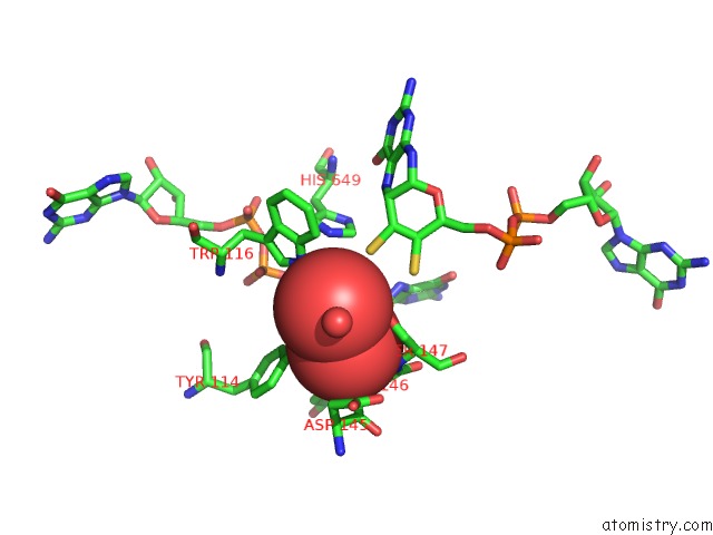
Mono view
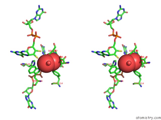
Stereo pair view
Mono view
Stereo pair view
A full contact list of Molybdenum with other atoms in the Mo binding
site number 1 of Structure of Dmso Reductase within 5.0Å range:
|
Reference:
F.Schneider,
J.Lowe,
R.Huber,
H.Schindelin,
C.Kisker,
J.Knablein.
Crystal Structure of Dimethyl Sulfoxide Reductase From Rhodobacter Capsulatus at 1.88 A Resolution. J.Mol.Biol. V. 263 53 1996.
ISSN: ISSN 0022-2836
PubMed: 8890912
DOI: 10.1006/JMBI.1996.0555
Page generated: Sun Oct 6 15:14:06 2024
ISSN: ISSN 0022-2836
PubMed: 8890912
DOI: 10.1006/JMBI.1996.0555
Last articles
Zn in 9MJ5Zn in 9HNW
Zn in 9G0L
Zn in 9FNE
Zn in 9DZN
Zn in 9E0I
Zn in 9D32
Zn in 9DAK
Zn in 8ZXC
Zn in 8ZUF