Molybdenum »
PDB 2ca3-3fah »
2dmr »
Molybdenum in PDB 2dmr: Dithionite Reduced Dmso Reductase From Rhodobacter Capsulatus
Protein crystallography data
The structure of Dithionite Reduced Dmso Reductase From Rhodobacter Capsulatus, PDB code: 2dmr
was solved by
A.S.Mcalpine,
S.Bailey,
with X-Ray Crystallography technique. A brief refinement statistics is given in the table below:
| Resolution Low / High (Å) | 20.00 / 2.80 |
| Space group | P 41 21 2 |
| Cell size a, b, c (Å), α, β, γ (°) | 81.030, 81.030, 230.040, 90.00, 90.00, 90.00 |
| R / Rfree (%) | 18.1 / 26.3 |
Molybdenum Binding Sites:
The binding sites of Molybdenum atom in the Dithionite Reduced Dmso Reductase From Rhodobacter Capsulatus
(pdb code 2dmr). This binding sites where shown within
5.0 Angstroms radius around Molybdenum atom.
In total only one binding site of Molybdenum was determined in the Dithionite Reduced Dmso Reductase From Rhodobacter Capsulatus, PDB code: 2dmr:
In total only one binding site of Molybdenum was determined in the Dithionite Reduced Dmso Reductase From Rhodobacter Capsulatus, PDB code: 2dmr:
Molybdenum binding site 1 out of 1 in 2dmr
Go back to
Molybdenum binding site 1 out
of 1 in the Dithionite Reduced Dmso Reductase From Rhodobacter Capsulatus
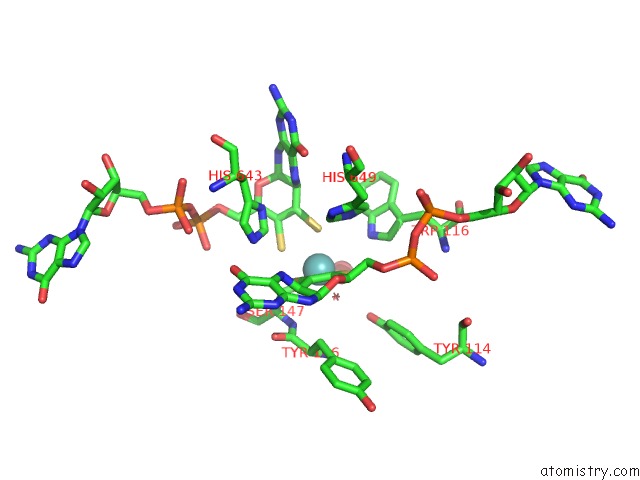
Mono view
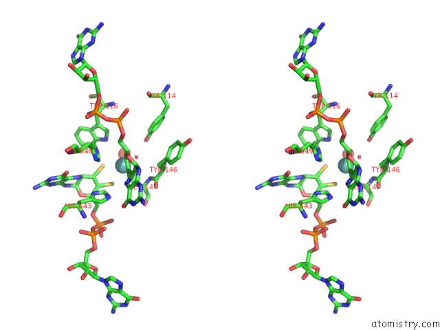
Stereo pair view
Mono view
Stereo pair view
A full contact list of Molybdenum with other atoms in the Mo binding
site number 1 of Dithionite Reduced Dmso Reductase From Rhodobacter Capsulatus within 5.0Å range:
|
Reference:
A.S.Mcalpine,
A.G.Mcewan,
A.Shaw,
S.Bailey.
Molybdenum Active Centre of Dmso Reductase From Rhodobacter Capsulatus: Crystal Structure of the Oxidised Enzyme at 1.82-A Resolution and the Dithionite-Reduced Enzyme at 2.8-A Resolution J.Biol.Inorg.Chem. V. 2 690 1997.
ISSN: ISSN 0949-8257
Page generated: Sun Oct 6 15:46:02 2024
ISSN: ISSN 0949-8257
Last articles
Mn in 6OIWMn in 6OI7
Mn in 6OIV
Mn in 6OE2
Mn in 6OBU
Mn in 6OBS
Mn in 6OBR
Mn in 6OBQ
Mn in 6O7E
Mn in 6O7D