Molybdenum »
PDB 3fc4-4c7z »
3hbg »
Molybdenum in PDB 3hbg: Structure of Recombinant Chicken Liver Sulfite Oxidase Mutant C185S
Protein crystallography data
The structure of Structure of Recombinant Chicken Liver Sulfite Oxidase Mutant C185S, PDB code: 3hbg
was solved by
J.A.Qiu,
with X-Ray Crystallography technique. A brief refinement statistics is given in the table below:
| Resolution Low / High (Å) | 27.38 / 1.90 |
| Space group | C 2 2 21 |
| Cell size a, b, c (Å), α, β, γ (°) | 86.468, 131.517, 98.884, 90.00, 90.00, 90.00 |
| R / Rfree (%) | 17.6 / 19.6 |
Molybdenum Binding Sites:
The binding sites of Molybdenum atom in the Structure of Recombinant Chicken Liver Sulfite Oxidase Mutant C185S
(pdb code 3hbg). This binding sites where shown within
5.0 Angstroms radius around Molybdenum atom.
In total only one binding site of Molybdenum was determined in the Structure of Recombinant Chicken Liver Sulfite Oxidase Mutant C185S, PDB code: 3hbg:
In total only one binding site of Molybdenum was determined in the Structure of Recombinant Chicken Liver Sulfite Oxidase Mutant C185S, PDB code: 3hbg:
Molybdenum binding site 1 out of 1 in 3hbg
Go back to
Molybdenum binding site 1 out
of 1 in the Structure of Recombinant Chicken Liver Sulfite Oxidase Mutant C185S
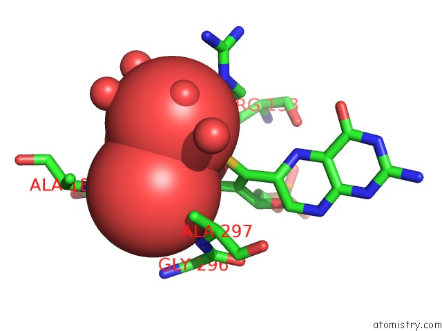
Mono view
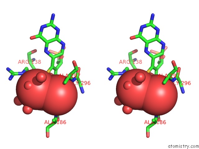
Stereo pair view
Mono view
Stereo pair view
A full contact list of Molybdenum with other atoms in the Mo binding
site number 1 of Structure of Recombinant Chicken Liver Sulfite Oxidase Mutant C185S within 5.0Å range:
|
Reference:
J.A.Qiu,
H.L.Wilson,
M.J.Pushie,
C.Kisker,
G.N.George,
K.V.Rajagopalan.
The Structures of the C185S and C185A Mutants of Sulfite Oxidase Reveal Rearrangement of the Active Site. Biochemistry V. 49 3989 2010.
ISSN: ISSN 0006-2960
PubMed: 20356030
DOI: 10.1021/BI1001954
Page generated: Sun Oct 6 15:53:24 2024
ISSN: ISSN 0006-2960
PubMed: 20356030
DOI: 10.1021/BI1001954
Last articles
K in 8XAKK in 8X9M
K in 8X70
K in 8WUW
K in 8WUX
K in 8X0S
K in 8X4R
K in 8WGR
K in 8W9V
K in 8W9O