Molybdenum »
PDB 6op4-8ccq »
7p41 »
Molybdenum in PDB 7p41: Crystal Structure of Human MARC1 A165T Variant
Enzymatic activity of Crystal Structure of Human MARC1 A165T Variant
All present enzymatic activity of Crystal Structure of Human MARC1 A165T Variant:
3.2.1.17;
3.2.1.17;
Protein crystallography data
The structure of Crystal Structure of Human MARC1 A165T Variant, PDB code: 7p41
was solved by
M.A.Struwe,
A.J.Scheidig,
with X-Ray Crystallography technique. A brief refinement statistics is given in the table below:
| Resolution Low / High (Å) | 31.92 / 1.60 |
| Space group | P 21 21 21 |
| Cell size a, b, c (Å), α, β, γ (°) | 61.063, 74.887, 111.164, 90, 90, 90 |
| R / Rfree (%) | 16 / 18.6 |
Other elements in 7p41:
The structure of Crystal Structure of Human MARC1 A165T Variant also contains other interesting chemical elements:
| Chlorine | (Cl) | 1 atom |
Molybdenum Binding Sites:
The binding sites of Molybdenum atom in the Crystal Structure of Human MARC1 A165T Variant
(pdb code 7p41). This binding sites where shown within
5.0 Angstroms radius around Molybdenum atom.
In total only one binding site of Molybdenum was determined in the Crystal Structure of Human MARC1 A165T Variant, PDB code: 7p41:
In total only one binding site of Molybdenum was determined in the Crystal Structure of Human MARC1 A165T Variant, PDB code: 7p41:
Molybdenum binding site 1 out of 1 in 7p41
Go back to
Molybdenum binding site 1 out
of 1 in the Crystal Structure of Human MARC1 A165T Variant
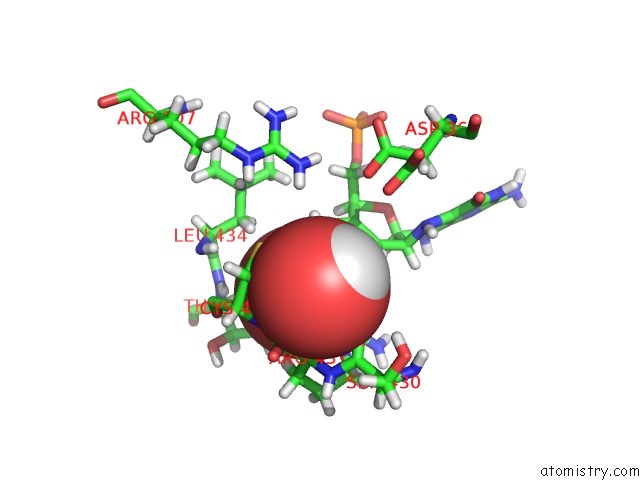
Mono view
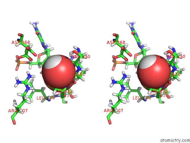
Stereo pair view
Mono view
Stereo pair view
A full contact list of Molybdenum with other atoms in the Mo binding
site number 1 of Crystal Structure of Human MARC1 A165T Variant within 5.0Å range:
|
Reference:
M.A.Struwe,
B.Clement,
A.J.Scheidig.
Crystal Structure of MARC1 A165T Variant at Near-Atomic Resolution To Be Published.
Page generated: Sun Oct 6 17:00:17 2024
Last articles
Mg in 7FJPMg in 7FQJ
Mg in 7FGI
Mg in 7FGG
Mg in 7FJJ
Mg in 7FH8
Mg in 7FH9
Mg in 7FH7
Mg in 7FH6
Mg in 7FH4