Molybdenum »
PDB 8cff-9cqy »
8rqz »
Molybdenum in PDB 8rqz: Crystal Structure of Molybdenum Bispyranopterin Guanine Dinucleotide Formate Dehydrogenases FORCE1 From Bacillus Subtilis
Protein crystallography data
The structure of Crystal Structure of Molybdenum Bispyranopterin Guanine Dinucleotide Formate Dehydrogenases FORCE1 From Bacillus Subtilis, PDB code: 8rqz
was solved by
M.V.Cherrier,
P.Arnoux,
L.Martin,
Y.Nicolet,
G.Schoehn,
P.Legrand,
M.Broc,
F.Seduk,
G.Brasseur,
R.Arias-Cartin,
A.Magalon,
A.Walburger,
A.Uzel,
B.Guigliarelli,
S.Grimaldi,
F.Pierrel,
M.Mate,
with X-Ray Crystallography technique. A brief refinement statistics is given in the table below:
| Resolution Low / High (Å) | 49.41 / 2.69 |
| Space group | F 2 2 2 |
| Cell size a, b, c (Å), α, β, γ (°) | 210.833, 210.5, 494.213, 90, 90, 90 |
| R / Rfree (%) | 19.6 / 23.1 |
Other elements in 8rqz:
The structure of Crystal Structure of Molybdenum Bispyranopterin Guanine Dinucleotide Formate Dehydrogenases FORCE1 From Bacillus Subtilis also contains other interesting chemical elements:
| Sodium | (Na) | 1 atom |
| Iron | (Fe) | 36 atoms |
Molybdenum Binding Sites:
The binding sites of Molybdenum atom in the Crystal Structure of Molybdenum Bispyranopterin Guanine Dinucleotide Formate Dehydrogenases FORCE1 From Bacillus Subtilis
(pdb code 8rqz). This binding sites where shown within
5.0 Angstroms radius around Molybdenum atom.
In total 2 binding sites of Molybdenum where determined in the Crystal Structure of Molybdenum Bispyranopterin Guanine Dinucleotide Formate Dehydrogenases FORCE1 From Bacillus Subtilis, PDB code: 8rqz:
Jump to Molybdenum binding site number: 1; 2;
In total 2 binding sites of Molybdenum where determined in the Crystal Structure of Molybdenum Bispyranopterin Guanine Dinucleotide Formate Dehydrogenases FORCE1 From Bacillus Subtilis, PDB code: 8rqz:
Jump to Molybdenum binding site number: 1; 2;
Molybdenum binding site 1 out of 2 in 8rqz
Go back to
Molybdenum binding site 1 out
of 2 in the Crystal Structure of Molybdenum Bispyranopterin Guanine Dinucleotide Formate Dehydrogenases FORCE1 From Bacillus Subtilis
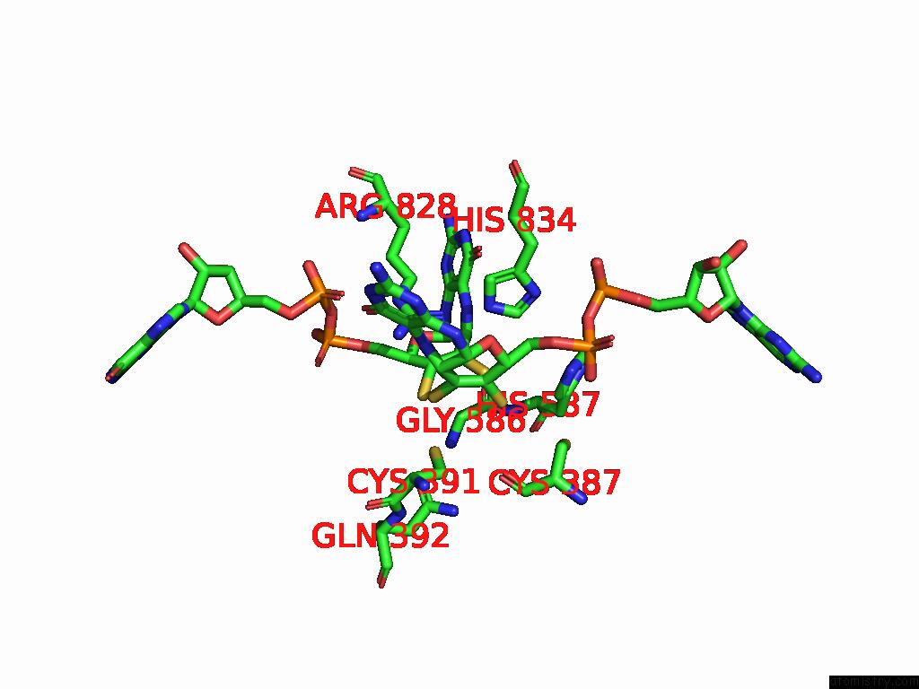
Mono view
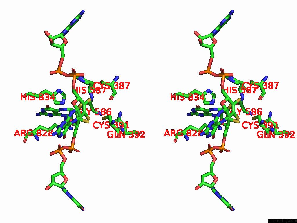
Stereo pair view
Mono view
Stereo pair view
A full contact list of Molybdenum with other atoms in the Mo binding
site number 1 of Crystal Structure of Molybdenum Bispyranopterin Guanine Dinucleotide Formate Dehydrogenases FORCE1 From Bacillus Subtilis within 5.0Å range:
|
Molybdenum binding site 2 out of 2 in 8rqz
Go back to
Molybdenum binding site 2 out
of 2 in the Crystal Structure of Molybdenum Bispyranopterin Guanine Dinucleotide Formate Dehydrogenases FORCE1 From Bacillus Subtilis
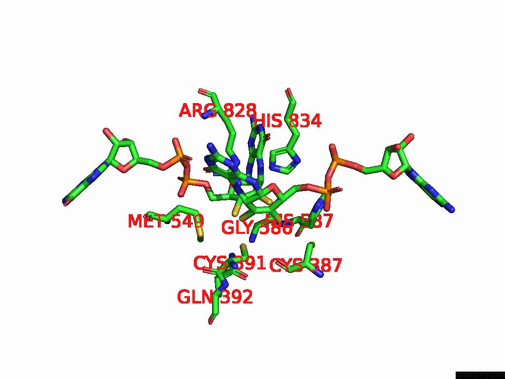
Mono view
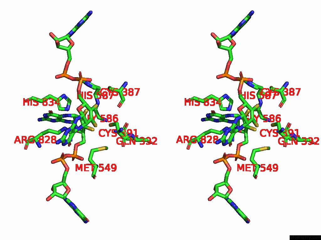
Stereo pair view
Mono view
Stereo pair view
A full contact list of Molybdenum with other atoms in the Mo binding
site number 2 of Crystal Structure of Molybdenum Bispyranopterin Guanine Dinucleotide Formate Dehydrogenases FORCE1 From Bacillus Subtilis within 5.0Å range:
|
Reference:
M.V.Cherrier,
P.Arnoux,
L.Martin,
Y.Nicolet,
G.Schoehn,
P.Legrand,
M.Broc,
F.Seduk,
G.Brasseur,
R.Arias-Cartin,
A.Magalon,
A.Walburger,
A.Uzel,
B.Guigliarelli,
S.Grimaldi,
F.Pierrel,
M.Mate.
Conserved Helical Membrane Plug-in For Quinone Reductases To Be Published.
Page generated: Sun Aug 17 04:34:49 2025
Last articles
Na in 4J4SNa in 4J9X
Na in 4J9W
Na in 4J9M
Na in 4J6W
Na in 4J4H
Na in 4J7V
Na in 4J4T
Na in 4J5Y
Na in 4J4B